Growth Potential and Management of Symptomatic Uterine Leiomyomas: Analysis of Two Clinical Cases
1Associate Professor A, EHS Nouar Fadéla, Oran, Algeria
2Associate Professor A, The Regional Military University Hospital of Oran, Algeria
3Professor and Head of Department, EHS Nouar Fadéla, Oran, Algeria
Author and article information
Cite this as
Sihem D, Skandar B, Yassine B. Growth Potential and Management of Symptomatic Uterine Leiomyomas: Analysis of Two Clinical Cases. Adv Women’s Health. 2026;4(1): 17-20. Available from: 10.17352/awh.000007
Copyright License
© 2026 Sihem D, et al. This is an open-access article distributed under the terms of the Creative Commons Attribution License, which permits unrestricted use, distribution, and reproduction in any medium, provided the original author and source are credited.Abstract
Uterine leiomyomas are the most common benign pelvic neoplasms in women of reproductive age. Although benign, specific subgroups exhibit aggressive growth dynamics leading to severe clinical complications, including acute anemia, pelvic pressure, and organ displacement [1]. This article presents a comparative analysis of two contrasting clinical cases of rapidly growing, highly symptomatic leiomyomas to highlight the key clinical drivers behind surgical decision-making and patient-tailored management.
Methods: We reviewed two anonymized medical records evaluating clinical presentation, preoperative optimization (specifically intravenous iron therapy), magnetic resonance imaging (MRI) mapping, surgical strategy (hysterectomy vs. myomectomy; ovarian preservation vs. bilateral salpingo-oophorectomy), and long-term postoperative outcomes.
Results: Case 1 presents a 45-year-old nulliparous patient with severe iron deficiency anemia (Hb 7.4 g/dL and a 25 cm confluent transmural leiomyoma mass. Despite nulliparity, total abdominal hysterectomy with ovarian preservation was performed due to total myometrial destruction, making polymyomectomy technically unfeasible. Case 2 involves a 49-year-old pauciparous patient with a 12 cm isthmic fibroid causing severe compressive urinary symptoms; total abdominal hysterectomy with bilateral salpingo-oophorectomy was selected given her perimenopausal age and involvement of adnexal structures. At 3-month and 6-month follow-ups, both patients achieved complete symptom resolution, with hemoglobin levels normalizing to 13.1 g/dL in Case 1.
Conclusion: Management of rapidly growing uterine fibroids requires individualized decision-making balancing tumor anatomy, clinical urgency, patient age, and fertility desires. Preoperative hemoglobin optimization and high-resolution MRI mapping are paramount to minimizing surgical morbidity and guiding optimal surgical approaches.
Introduction
Uterine leiomyomas, commonly known as fibroids, represent the most frequent benign pelvic tumors in women of childbearing age, with an estimated prevalence ranging from 70% to 80% according to histological data. Although essentially benign, these monoclonal tumors originating from the myometrium can exhibit marked growth dynamics driven by hormonal factors (estrogens and progesterone) and complex molecular signaling pathways (notably the extracellular matrix and growth factors) [1].
Clinically, certain fibroids are distinguished by a high growth potential and debilitating symptoms (severe menorrhagia, anemia, pelvic pain, and pelvic static disorders). This article aims to illustrate the variability of clinical presentation and therapeutic strategies through the analysis of two clinical cases of patients presenting with rapidly growing leiomyomas [2].
Methods
This is a retrospective analysis of two anonymized medical records of patients treated for symptomatic uterine leiomyomas characterized by rapid growth dynamics at EHS Nouar Fadéla University Hospital of Oran.
Evaluated parameters
- Clinical evaluation: Functional symptoms (menorrhagia, dysmenorrhea, urinary frequency), symptom duration, impact on quality of life, and gynecological history.
- Medical imaging: High-resolution pelvic ultrasonography and pelvic Magnetic Resonance Imaging (MRI) to assess uterine volume, leiomyoma count, FIGO classification, exact spatial localization, and proximity to pelvic organs.
- Biological parameters: Serial hemoglobin (Hb) levels, serum ferritin, and coagulation profile.
- Therapeutic strategy: Rationale behind surgical selection (conservative myomectomy vs. radical hysterectomy; adnexal preservation vs. bilateral salpingo-oophorectomy), preoperative optimization, and discussion of alternative non-invasive modalities.
- Post-therapeutic outcomes: Immediate postoperative course, wound healing, serial hematological recovery at 3 months, and complete clinical resolution at 6 months.
Case Report 1
A 45-Year-Old Patient with Multiple Rapidly Growing Submucosal and Intramural Fibroids, Acute Anemia, and Mass Effect
Clinical presentation
A 45-year-old nulliparous patient presented with debilitating, heavy menorrhagia ongoing for six months, associated with a rapid increase in abdominal volume, secondary dysmenorrhea, and severe fatigue. Clinical examination revealed a mass inseparable from the uterus, reaching the size of a 6-month pregnancy. Laboratory tests showed severe iron deficiency anemia (hemoglobin level at 7.4 g/dL).
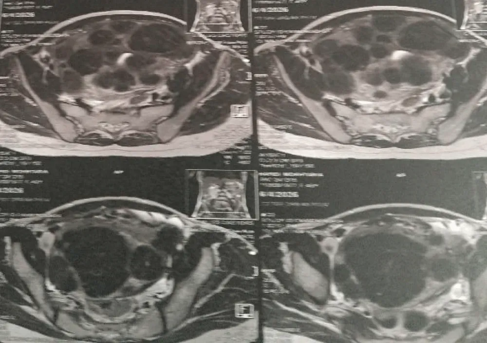
Imaging: Pelvic ultrasound and MRI demonstrated an enlarged uterus with multiple confluent FIGO-type leiomyomas (intracavitary submucosal and intramural) measuring 25 cm in maximum diameter, characterized by a rapid expansion of abdominal abdominal volume compared to previous months [1] (Figure 1).
Surgical justification and alternative
Treatments: Alternative uterine-preserving or minimally invasive modalities (such as Uterine Artery Embolization [UAE], High-Intensity Focused Ultrasound [HIFU], or medical therapy with GnRH agonists/antagonists) were thoroughly evaluated and discussed with the patient. GnRH agonists were deemed insufficient as sole therapy given the massive 25 cm tumor burden and acute clinical deterioration. UAE and HIFU were ruled out due to the high risk of incomplete necrosis, transvaginal expulsion of large submucosal fragments, secondary infection, and the urgent need for mass relief.
Regarding surgical approach: Although the patient was nulliparous, organ-preserving polymyomectomy was technically unfeasible due to total structural destruction of the surrounding myometrial tissue and the extreme risk of uncontrollable intraoperative hemorrhage under general anesthesia. Following an extensive multidisciplinary board review and informed patient consent, a Total Abdominal Hysterectomy with Ovarian Preservation was elected. Preserving the ovaries was crucial to avoid premature surgical menopause and maintain endogenous estrogenic support in this 45-year-old patient.
Management and surgical outcome
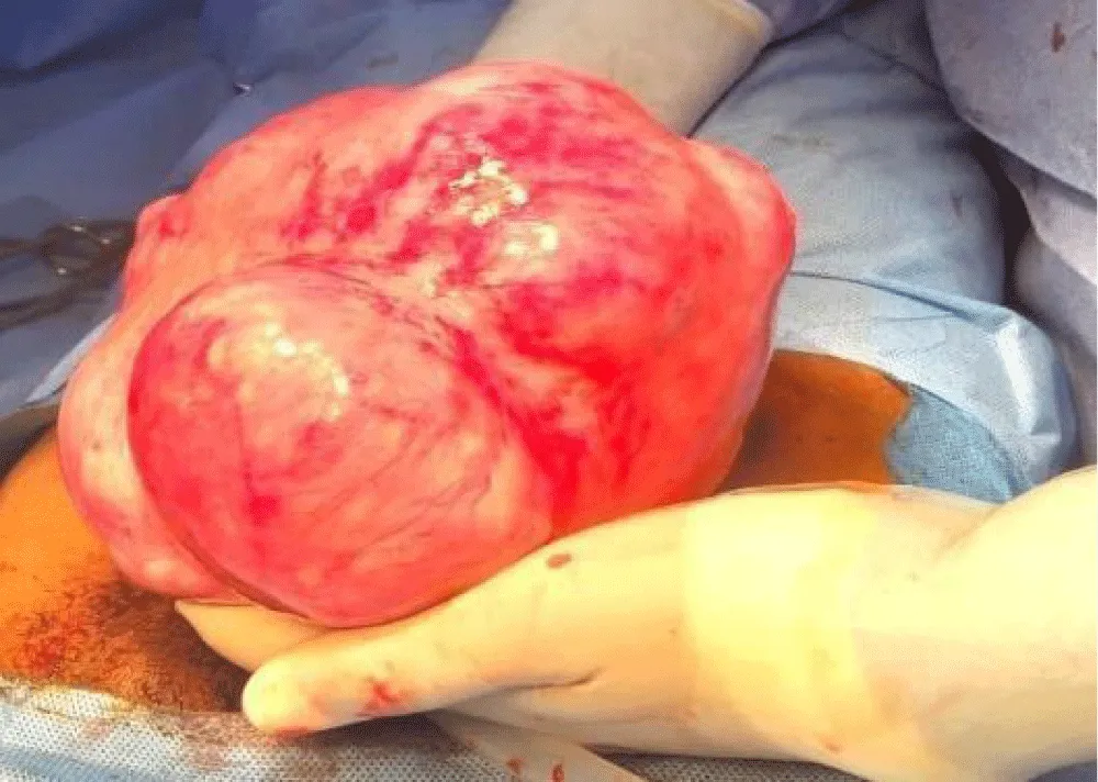
Preoperative optimization was initiated with intravenous iron sucrose infusions, raising the hemoglobin level from 7.4 g/dL to 10.2 g/dL prior to surgery, avoiding the necessity of homologous blood transfusion. Total abdominal hysterectomy was performed successfully.
Figure 2: Intraoperative view showing surgical exploration of the massive uterine tumor (25 cm) occupying the abdominopelvic cavity before resection.
Follow-up and outcomes
The immediate postoperative period was uneventful. At the 3-month follow-up, the patient exhibited excellent surgical wound healing and complete hematological recovery, with a hemoglobin level reaching 13.1g/dL. At the 6-month follow-up, the patient reported complete resolution of fatigue and pelvic discomfort, with a normal hormonal profile and no surgical sequelae.
Case Report 2
A 49-Year-Old Patient with Large Isthmic Fibroid and Compression Syndrome:
Clinical presentation: A 49-year-old pauciparous patient presented with a progressive and rapid increase in abdominal volume over the past twelve months, associated with urinary frequency (compressive urinary symptoms) and chronic pelvic heaviness.
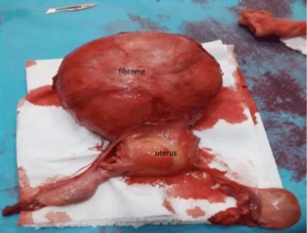
Imaging: Pelvic MRI revealed a massive multinodular uterus with three large leiomyomas (including a 12 cm isthmic fibroid with a subserosal component), altering the axis of the uterine cavity and compressing the posterior wall of the bladder (Figure 3).
Surgical justification and alternative treatments: Given the patient’s age (49 years, perimenopausal state) and persistent lower urinary tract symptoms, conservative medical management (such as GnRH receptor antagonists) offered only temporary relief with high symptom relapse rates upon cessation. Uterine Artery
Embolization was discussed but declined by the patient in favor of a definitive surgical solution.
Total Abdominal Hysterectomy with Bilateral Salpingo-Oophorectomy (TAH-BSO) was selected. Hysterectomy was indicated due to the anatomical location of the 12 cm isthmic fibroid, which effaced the lower uterine segment, making isolated myomectomy technically treacherous with high risks of cervical injury and severe hemorrhage. Bilateral salpingo-oophorectomy was performed following thorough preoperative discussion, taking into account her perimenopausal age (49 years), complete family planning, and the clinical intent to eliminate the risk of subsequent ovarian and fallopian tube malignancies, aligning with international gynecological standards for perimenopausal hysterectomy.
Follow-up and outcomes
Postoperative recovery was rapid and uneventful. Compressive lower urinary tract symptoms (pollakiuria and bladder pressure) resolved completely within two weeks post-surgery. Histopathological analysis confirmed benign uterine leiomyomas without cellular atypia, pleomorphism, or coagulative tumor necrosis. At the 6-month follow-up evaluation, the patient remained fully asymptomatic with no urinary dysfunction or surgical complications.
Discussion
Uterine leiomyomas are estrogen- and progesterone-dependent tumors, whose rapid development can be explained by complex interactions involving steroid hormones, local growth factors, and alterations in the extracellular matrix. Although the vast majority of fibroids exhibit slow growth, a subgroup shows high proliferative dynamics, posing significant diagnostic and therapeutic challenges.
Mass effect and compressive symptoms
Large leiomyomas, particularly those located in the isthmus or lower uterine segment, can cause severe compressive symptoms on adjacent pelvic structures (bladder, ureters, rectum). As illustrated in Case 2, subserosal and isthmic development directly impacted the bladder, leading to urinary frequency and pelvic pressure.
1. Hematologic Impact:
Intramural and submucosal fibroids frequently cause severe menorrhagia, leading to chronic or acute iron deficiency anemia (as observed in Case 1 with a hemoglobin level of 7.4 g/dL). Preoperative optimization, including intravenous iron therapy, is essential to decrease surgical morbidity and avoid homologous blood transfusions.
2. Surgical Strategy:
The management of rapidly growing or large symptomatic leiomyomas must be tailored to the patient’s age, desire for future fertility, symptom severity, and tumor location. While conservative treatment (myomectomy) is preferred in women wishing to preserve fertility, radical surgery (hysterectomy) remains the definitive treatment option in peri-menopausal patients or when structural uterine destruction renders organ-sparing surgery unfeasible [3-5].
Comparison of treatment strategies
Limitations
This study is limited by its retrospective, two-case design, which precludes broad epidemiological generalizations. Additionally, long-term molecular profiling (e.g., MED12 mutation status or HMGA2 overexpression) was not performed on the surgical specimens, which could have provided deeper mechanistic insights into their rapid growth dynamics.
Conclusion
Symptomatic uterine leiomyomas with high growth dynamics require precise clinical and radiological evaluation to guide therapeutic decision-making. Pelvic MRI plays a crucial role in mapping the lesions and planning the surgical approach. A multidisciplinary management plan, combining preoperative hematologic optimization and tailored surgical techniques, allows for successful symptom relief and favorable postoperative outcomes.
References
- Stewart EA, Cookson CL, Gandolfo RA, Schulze-Rath R. Epidemiology of uterine fibroids: a systematic review. BJOG. 2017;124(10): 1501-1512. Available from: https://doi.org/10.1111/1471-0528.14640
- Bulun SE. Uterine fibroids. N Engl J Med. 2013:369(14); 1344-1355. Available from: https://doi.org/10.1056/nejmra1209993
- Donnez J, Dolmans MM. Uterine fibroid management: from the past to the future. Hum Reprod Update. 2016; 22(6): 665-686. Available from: https://doi.org/10.1093/humupd/dmw023
- Parker WH. Etiology, symptomatology, and diagnosis of uterine myomas. Fertil Steril. 2007;87(4): 725-736. Available from: https://doi.org/10.1016/j.fertnstert.2007.01.093
- Stewart EA. Uterine fibroids. Lancet. 2015;385(9978): 1630-1640.
Advertisement

 Save to Mendeley