Global Journal of Medical and Clinical Case Reports
Aggressive Angiomyxoma of the Cervix: A Unique Entity
1Obstetrics & Gynecology, PGIMER, Chandigarh, India
2Department of Pathology, Government Medical College & Hospital, Chandigarh, India
3Obstetrics & Gynecology, Government Medical College & Hospital, Chandigarh, India
Author and article information
Cite this as
Siwatch S, Kundu R, Takkar N (2014) Aggressive Angiomyxoma of the Cervix: A Unique Entity. Global J Med Clin Case Reports 1(1): 010-012. DOI: 10.17352/2455-5282.000003
Copyright License
© 2014 Siwatch S, et al. This is an open-access article distributed under the terms of the Creative Commons Attribution License, which permits unrestricted use, distribution, and reproduction in any medium, provided the original author and source are credited.Abstract
Introduction: Aggressive angiomyxoma is a mesenchymal tumour which presents rarely; usually in women in reproductive age group, as a mass in the pelvic tissue. Only exceptionally, has it been seen to originate from the uterus and cervix.
Methods: We present a case of a 23-year-old woman who complained about abdominal pain and excessive vaginal bleeding three days after a spontaneous abortion. Examination revealed a large polyp arising from the cervix. Histopathologic examination of the polypectomy specimen revealed an aggressive angiomyxoma of the cervix. Immunohistochemistry was performed and the tumour was immunopositive for vimentin, desmin and smooth muscle actin. Management options are discussed, in light of need of fertility conservation.
Result: Aggressive angiomyxoma can originate from the mesenchymal tissue of the cervix.
Conclusions: Treatment may be individualized with surgery and hormonal treatment. Long term follow up is recommended.
Introduction
Aggressive angiomyxoma is a rare locally invasive soft tissue tumour arising from the perineum. First reported by Steeper and Rosai in 1983, it is usually asymptomatic and presents as a mass in the perineum [1]. Aggressive angiomyxoma is thought to be estrogen progesterone sensitive and occurs usually in the reproductive age group [2]. Here, we present a case with an uncommon origin of this rare tumour.
Case Presentation
A 23-year-old woman presented to the outpatient clinic with complaints of excessive vaginal bleeding for three days after an abortion. It was her first pregnancy and she aborted spontaneously at home. She had a four month period of amenorrhea but a pregnancy test or ultrasound had not been done. Previous menstrual cycles were normal and regular. General examination was unremarkable except for evidence of anemia. Pelvic examination revealed a large fleshy polypoidal mass protruding from the cervix which was felt high up in the vagina. Though the patient did have some dyspareunia and vaginal discharge, there was no history of post coital bleeding.
Urine pregnancy test was negative. Ultrasound showed a large 8 cm x 6 cm x 5 cm fibroid polyp in the cervix. The differential diagnoses of a cervical polyp or a retained placental polyp after spontaneous abortion were considered. The lady underwent removal of the cervical polyp under spinal anaesthesia. The cervix was ballooned and dilated. The large fleshy polyp was divided into 2 parts for removal. The blood loss was approximately 20 ml. The specimen was sent for histopathology (Figure 1).
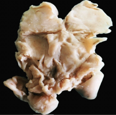
Gross specimen showing bisected polyp with homogeneously smooth and glistening sectioned surface.
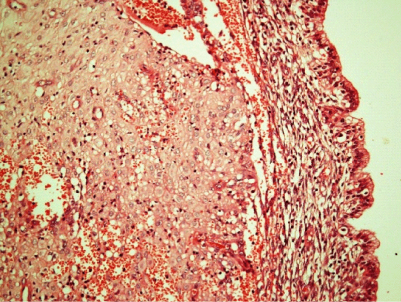
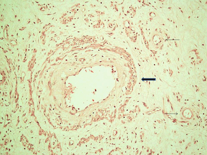
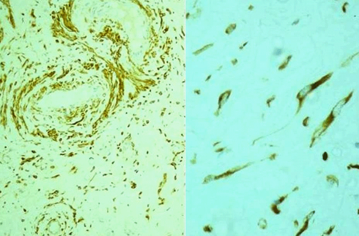
The patient was discharged in a satisfactory condition on post-operative day three. The histopathology of the cervical polyp (Figure 2) unexpectedly reported low cellularity with scattered spindle cells in a myxoid background; features suggestive of aggressive angiomyxoma (AAM) of the cervix (Figure 3). The immunohistochemistry was positive for smooth muscle actin (SMA), vimentin and desmin while S-100 was negative (Figure 4). The excisional margins were free of tumor and the surgical treatment was complete. However, aggressive angiomyxoma has a high rate of local recurrence and long term follow up is required which in our case would include clinical symptomatology, physical examination and imaging with either ultrasound and/ or magnetic resonance imaging (MRI) of the pelvis to evaluate the uterus and adnexa. In case of recurrence, a hysterectomy may be required to completely remove the local uterine disease. All the same, our patient is since lost to follow up and has not reported back in the last 2 years.
Discussion
Aggressive angiomyxoma is a rare mesenchymal tumour that presents to the gynecologists as a growth/ mass arising from the vulva or perineum; mistaken as a lipoma, bartholin, gartner's cyst or labial mass and has no typical symptoms [2-4]. Patients are often asymptomatic and compression to urinary/intestinal systems occurs only when the tumour is large. Very rarely, and to our knowledge, only two cases have been reported where it has presented as a cervical polyp with symptoms of distension or a uterine polyp where it presented with uterine bleeding and urinary retention [5,6]. Aggressive angiomyxoma is thought to be estrogen and progesterone sensitive and presents usually in the reproductive age group, with some cases reporting growth of the tumour during pregnancy [2,7]. The elevated hormone levels during the antecedent pregnancy may have contributed to growth of the tumour in our patient too. However, the peculiar location of the tumour in our patient probably led to the dilation of the cervix that led to the spontaneous abortion in this patient.
An accurate diagnosis of aggressive angiomyxoma is based on histopathology evidence. Grossly these tumours have a smooth surface with homogenous consistency and a glistening gelatinous appearance on cut section [1]. Microscopy shows a hypocellular mesenchymal lesion with scattered bland spindled and stellate cells in a myxoid stroma. The tumour lacks cytologic atypia, increased/atypical mitoses and coagulative necrosis. Characteristic are the numerous blood vessels of varying calibre as seen in the current case. The tumour is usually positive for vimentin, desmin and negative for S-100 on imomunohistochemistry [8]. However, this immunoprofile is shared by superficial cervicovaginal myofibroblastoma [9]. Sparse cellularity, more infiltrative pattern and variability in distribution, size and wall thickness of vessels favour aggressive angiomyxoma.
Aggressive angiomyxoma is rare and usually presents with a slow growing mass due to which it may be mistaken for a benign growth. Pathologically, aggressive angiomyxoma needs to be differentiated from angiomyofibroblastoma (AMF), myxoid leiomyoma, cellular angiofibroma, superficial myofibroblastoma, myxoid liposarcoma and other differentials that may mimic aggressive angiomyxoma and differ in the management, thus emphasizing the need for pre-operative diagnosis [7,10]. Prior to definitive management, a pre-operative fine needle aspiration (FNA) may help exclude lymphoproliferative and metastatic pathologies [10]. Radiological assessment of extent of lesion using radiological modalities like ultrasound, computerized tomography (CT) and MRI should be considered to help plan surgery [7,11]. On CT, it is seen as a well-defined mass, hypodense as compared to the muscle. AAM is seen as a hypointense lesion on T1 weighted MRI images and hyperintense lesion on T2 weighted images [11].
Angiomyxoma is thought to displace rather than invade surrounding tissue. In the usual case of AAM presenting as a perineal mass, careful anatomic dissection should be done to avoid injury to anal sphincters and lower urinary system that may be in close proximity in an AAM [2,4]. In case of a cervical or uterine polyp, the complete removal of the polyp and the pedicle should be done to ensure disease free margins, which should be confirmed by histopathology, as in our case. AAM has high recurrence (36-72%), is locally invasive and few metastasis have been reported [2,3,11,12]. Surgery with wide excision and tumour free margins has been advocated [2-4]. However, the chances of local recurrence are similar with complete tumour free margins and without [2,11]. Follow up assessment of treatment response should include clinical examination and CT/ MRI [2]. Our patient was planned for follow up with CT/ MRI to assess for recurrence, especially in the cervix, uterus and pelvis. In case of involvement of the cervix/ uterus, local resection would warrant a hysterectomy.
Other therapeutic options could be used to preserve fertility. These include hormonal treatment with gonadotropin releasing hormone (GnRH) agonists or tamoxifen [3,11,13]. Leuprolide acetate administered intramuscularly in monthly doses of 3.75 mg has been used [14]. Tamoxifen has also been used in the treatment of aggressive angiomyxoma due to its antiestrogenic action [12]. These agents may help in reducing the extent of surgery by reducing the size of the tumour [11]. Even though radiotherapy and chemotherapy may seem to be less useful due to the low mitotic activity and low cellularity of the tumour, various reports have documented reduced recurrence with their use [7,15]. The presence of multiple feeding vessels also limits the use of embolization as a treatment modality [11]. Individualization of the treatment options and multimodal treatment seem to be the most appropriate [16,17].
Conclusions
Aggressive angiomyxoma should be considered in the differential diagnosis of a uterine or cervical polyp, especially when presenting in pregnancy. When presenting as a polyp, aggressive angiomyxoma could lead to spontaneous abortion. Treatment options include surgical resection and hormonal therapy like GnRH agonists and tamoxifen. In case of uterine/ cervical polyp, reappearance of symptomatology, ultrasound, CT/ MRI and curettage may be used to assess for recurrence. In case of recurrence of angiomyxoma, local resection may require hysterectomy. Conservative options in the form of GnRH agonists and tamoxifen may be offered to preserve fertility. Long term follow up is recommended.
References
- Steeper TA, Rosai J (1983) Aggressive angiomyxoma of the female pelvis and perineum. Report of nine cases of a distinctive type of gynecologic soft-tissue neoplasm. Am J Surg Pathol 7: 463-475.
- Chan YM, Hon E, Ngai SW, Ng TY, Wong LC (2000) Aggressive angiomyxoma in females: is radical resection the only option? Acta Obstet Gynecol Scand 79: 216-220.
- Han-Geurts IJ, van Geel AN, van Doorn L, den Bakker M, Eggermont AM, et al. (2006) Aggressive angiomyxoma: multimodality treatments can avoid mutilating surgery. Eur J Surg Oncol 32: 1217-1221.
- Behranwala KA, Thomas JM (2003) ‘Aggressive' angiomyxoma: a distinct clinical entity. Eur J Surg Oncol 29: 559-563.
- Yaghoobian J, Zinn D, Ramanathan K, Pinck RL, Hilfer J (1987) Ultrasound and computed tomographic findings in aggressive angiomyxoma of the uterine cervix. JUM 6: 209-212.
- Skálová A, Zámecník M, Michal M, Opatrny V (2000) Aggressive angiomyxoma presenting as polyp of uterine cavity. Pathol Res Pract 196: 719-725.
- Bagga R, Keepanasseril A, Suri V, Nijhawan R (2007) Aggressive angiomyxoma of the vulva in pregnancy: a case report and review of management options. Med GenMed 9:16.
- Zhang P, Song K, Li L, Geng F, Li J, et al. (2011) Aggressive angiomyxoma with massive ascites. Gynecol Oncol Case Rep 1: 12-13.
- Laskin WB, Fetsch JF, Tavassoli FA (2001) Superficial cervicovaginal myofibroblastoma: fourteen cases of a distinctive mesenchymal tumor arising from the specialized subepithelial stroma of the lower female genital tract. Hum Pathol 32: 715-725.
- Layfield LJ, Dodd LG (1997) Fine-needle aspiration cytology findings in a case of aggressive angiomyxoma: a case report and review of the literature. Diagn Cytopathol 16: 425-429.
- Haldar K, Martinek IE, Kehoe S (2010) Aggressive angiomyxoma: a case series and literature review. Eur J Surg Oncol 36: 335-339.
- Tsarpalis DC, Giannakopoulos CK, Lagadas AA, Toufexi EG (2007) Aggressive angiomyxoma: case report and review of the literature. Eur J Gynaecol Oncol 28: 480-482.
- Lee CW, Yoon JH, Park DC, Lee SJ (2011) Aggressive angiomyxoma of the vulva treated by using a gonadotropin-releasing hormone agonist: a case report. Eur J Gynaecol Oncol 32: 686-688.
- Poirier M, Fraser R, Meterissian S (2003) Case 1. Aggressive angiomyxoma of the pelvis: response to luteinizing hormone-releasing hormone agonist. J Clin Oncol 21: 3535-3536.
- Rhomberg W, Jasarevic Z, Alton R, Kompatscher P, Beer G, et al. (2000) Aggressive angiomyxoma: irradiation for recurrent disease. Strahlenther Onkol 176: 324-326.
- Coppola S, Desai A, Tzanis D, Honoré C, Bitsakou G, et al. (2013) Conservative en bloc surgery for aggressive angiomyxoma achieves good local control: analysis of 14 patients from a single institution. Int J Gynecol Cancer 23: 540-545.
- Bai HM, Yang JX, Huang HF, Cao DY, Chen J, et al. (2013) Individualized managing strategies of aggressive angiomyxoma of female genital tract and pelvis. Eur J Surg Oncol 39: 1101-1108.
Advertisement