Global Journal of Medical and Clinical Case Reports
Case Report of Mammary-Type Myofibroblastoma with Sarcomatous Features
1Megalab- Medical Laboratory in Georgia, Tbilisi, Georgia
2Labpon-Laboratory Pathology, Netherlands
3Davit Tvildiani Medical University, Tbilisi, Georgia
Author and article information
Cite this as
Tugushi D, Vink R, Baidoshvili A. Case Report of Mammary-Type Myofibroblastoma with Sarcomatous Features. Glob J Medical Clin Case Rep. 2026:13(7):138-141. Available from: 10.17352/gjmccr.000258
Copyright License
© 2026 Tugushi D, et al. This is an open-access article distributed under the terms of the Creative Commons Attribution License, which permits unrestricted use, distribution, and reproduction in any medium, provided the original author and source are credited.Abstract
Mammary-type Myofibroblastoma (MFB) is a rare benign mesenchymal neoplasm that most commonly occurs in the breast and along the embryonic milk line. Histologically, it is characterised by spindle-shaped cells with myofibroblastic differentiation and typically demonstrates an indolent clinical course. Although several histological variants have been described, tumours exhibiting atypical or sarcomatous-like features are exceptionally uncommon. We report the case of a 78-year-old woman who presented with a slowly enlarging right breast mass. Core needle biopsies performed one year apart demonstrated the characteristic morphology and immunophenotype of mammary-type myofibroblastoma. Two years later, following interval enlargement of the lesion, lumpectomy revealed a biphasic tumour composed of a conventional mammary-type myofibroblastoma and a sharply demarcated atypical component exhibiting increased cellularity, marked nuclear pleomorphism, hyperchromasia, multinucleation, and increased mitotic activity. Immunohistochemical analysis showed diffuse expression of desmin, Estrogen Receptor (ER), Androgen Receptor (AR), and Smooth Muscle Actin (SMA) in both components. In contrast, the atypical component demonstrated reduced CD34 expression, loss of progesterone receptor (PR) expression, increased Ki-67 proliferative activity, and diffuse p53 positivity. These immunohistochemical alterations were confined to the atypical area and may suggest biological progression; however, molecular genetic studies would be required to establish a clonal relationship between the conventional and atypical components. This case expands the limited literature on mammary-type myofibroblastoma with sarcomatous-like features and emphasises the importance of recognising this rare morphological variant to avoid diagnostic pitfalls and unnecessary overtreatment.
Introduction
Mammary-type myofibroblastoma (MFB) is a rare benign mesenchymal neoplasm composed of spindle cells with myofibroblastic differentiation. It was first described by Wargotz et al. in 1987 as a distinctive benign stromal tumour of the breast. Since then, morphologically identical tumours have been reported in a variety of extramammary locations along the embryonic milk line as well as in other soft tissues [1,2].
Clinically, MFB usually presents as a slow-growing, painless, well-circumscribed, mobile breast mass. Imaging findings are nonspecific, and the lesion may mimic other benign or malignant spindle cell tumours. Histopathological examination, supported by immunohistochemistry, is therefore essential for establishing the correct diagnosis. Typical tumours show diffuse expression of CD34, desmin, Estrogen Receptor (ER), Androgen Receptor (AR), and Smooth Muscle Actin (SMA), together with deletion of the 13q14 region, supporting its relationship with spindle cell lipoma and cellular angiofibroma [2,3].
Although MFB is regarded as a benign neoplasm with an excellent prognosis following complete surgical excision, rare cases with atypical or sarcomatous-like features have been described. We report an unusual case of mammary-type myofibroblastoma showing focal sarcomatous-like transformation and discuss its histopathological and immunohistochemical characteristics in the context of the current literature [4,5].
Case presentation
A 78-year-old woman presented with a palpable mass in the upper outer quadrant of the right breast. The lesion had been slowly increasing in size over approximately two years. Mammography and ultrasonography demonstrated a well-circumscribed solid mass measuring 24 × 13 mm. No axillary lymphadenopathy or other suspicious lesions were identified.
An ultrasound-guided core needle biopsy was initially performed. Histopathological examination revealed a spindle cell lesion composed of bland spindle-shaped cells arranged in short intersecting fascicles separated by hyalinized collagen bundles. No significant cytologic atypia, necrosis, or increased mitotic activity was identified.
One year later, because of interval enlargement of the lesion, a repeat core needle biopsy was performed and demonstrated similar morphological findings. Owing to the continued increase in tumour size, breast-conserving surgical excision (lumpectomy) was subsequently undertaken.
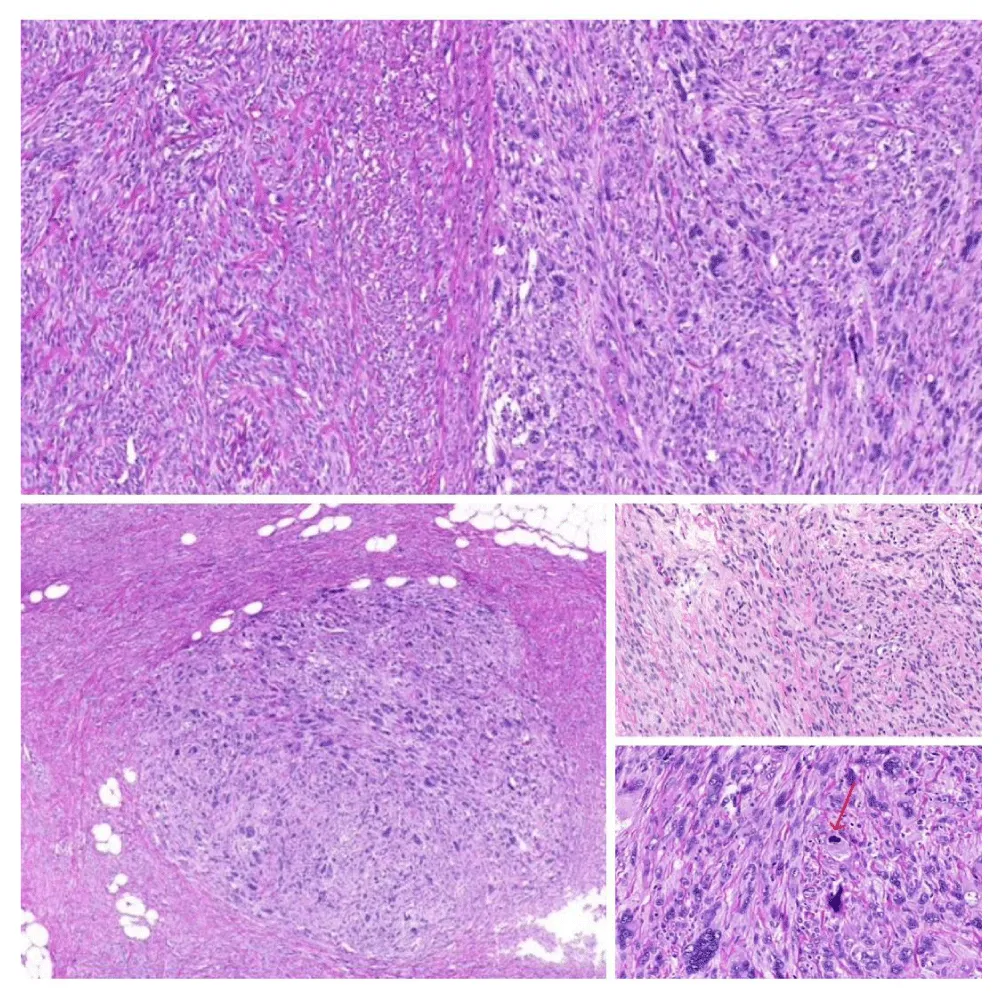
Gross examination revealed a well-circumscribed, firm, gray-white mass measuring 24 × 13 mm. Microscopically, most of the tumour exhibited the characteristic morphology of mammary-type myofibroblastoma. However, a sharply demarcated focal area demonstrated increased cellularity, marked nuclear pleomorphism, hyperchromasia, multinucleation, and increased mitotic activity, consistent with sarcomatous-like transformation.
Results
Histological and Immunohistochemical Findings
Both initial core needle biopsy specimens demonstrated the characteristic morphology of mammary-type myofibroblastoma, consisting of bland spindle cells arranged in short fascicles within a hyalinized collagenous stroma. No mitotic activity or necrosis was identified. Immunohistochemical analysis revealed diffuse positivity for desmin, Estrogen Receptor (ER), Androgen Receptor (AR), and CD34. Progesterone Receptor (PR) showed focal weak positivity, and the Ki-67 labelling index was approximately 5%. Cytokeratin 8/18 (CK8/18), AE1/AE3, and Retinoblastoma Protein (Rb) were negative.
Histological examination of the excised 24 × 13 mm lesion demonstrated a biphasic tumour. The predominant component exhibited the characteristic features of mammary-type myofibroblastoma, composed of spindle cells with oval nuclei, inconspicuous nucleoli, and abundant eosinophilic collagenous stroma. In contrast, a sharply demarcated atypical component demonstrated increased cellularity, marked nuclear pleomorphism, hyperchromasia, multinucleation, reduced stromal collagen, and occasional mitotic figures. The atypical cells were arranged in a haphazard fascicular pattern and showed focal infiltration into the surrounding adipose tissue.
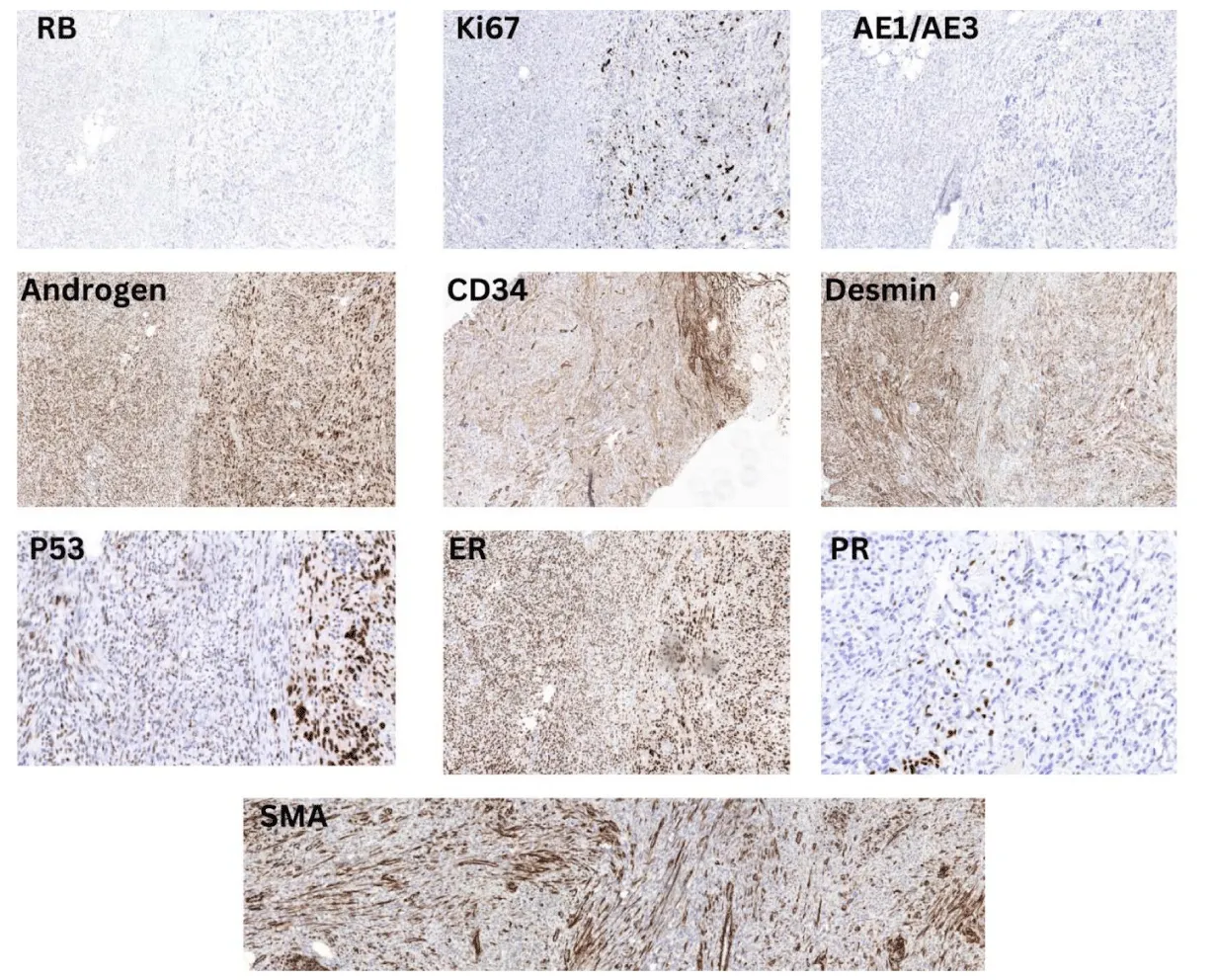
Immunohistochemical analysis demonstrated reduced CD34 expression in the atypical component compared with the conventional component. Desmin, Estrogen Receptor (ER), Androgen Receptor (AR), and Smooth Muscle Actin (SMA) remained diffusely positive in both tumour components. Progesterone Receptor (PR) expression was absent in the atypical component, whereas the Ki-67 labelling index was markedly increased. Diffuse p53 positivity was observed exclusively in the atypical component, while the conventional component remained negative. These immunohistochemical findings were confined to the histologically atypical area of the tumour.
Discussion
Mammary-type Myofibroblastoma (MFB) is a rare benign mesenchymal tumour with a well-documented indolent clinical course and an excellent prognosis following complete surgical excision. Although several histological variants have been described, tumours exhibiting atypical or sarcomatous-like features remain exceptionally uncommon [1,2,4].
The present case demonstrated an abrupt transition between a conventional MFB component and an atypical component characterised by increased cellularity, nuclear pleomorphism, hyperchromasia, multinucleation, and increased mitotic activity. These morphological features closely resemble the atypical or sarcomatous changes previously reported in cellular angiofibroma, supporting the concept that these tumours may share overlapping histopathological characteristics [6,7]. Cellular angiofibroma and mammary-type myofibroblastoma also share several morphological, immunophenotypic, and genetic features, including frequent CD34 expression and deletion of the 13q14 region, suggesting a close biological relationship between these entities [3,8].
In the present case, the atypical component demonstrated reduced CD34 expression, loss of progesterone receptor (PR) expression, increased Ki-67 proliferative activity, and diffuse p53 positivity. These findings were confined to the morphologically atypical area of the tumour. Although these immunohistochemical alterations may indicate biological progression, they are insufficient to establish a clonal relationship between the conventional and atypical components without molecular genetic analysis.
In comparison with previously reported cases of mammary-type myofibroblastoma with sarcomatous or atypical features, the present case shares several important clinical, histopathological, and immunohistochemical characteristics [2,4,5]. Similar to previously reported patients, our patient presented with a slowly enlarging, well-circumscribed breast mass and underwent complete surgical excision as definitive treatment [2,9]. Histologically, the tumour exhibited the characteristic spindle cell morphology and diffuse expression of CD34 and desmin in the conventional component, consistent with previous reports [2,4]. However, unlike most published cases, our tumour demonstrated a focal area of abrupt sarcomatous-like transformation characterised by marked nuclear atypia, pleomorphism, increased mitotic activity, and reduced stromal collagen [4,5]. Such changes have only rarely been described in the literature and may represent a possible spectrum of tumour progression rather than unequivocal malignant transformation [4,7]. Nevertheless, molecular studies are required to determine whether the conventional and atypical components are clonally related. Although the available literature indicates that most patients experience a favourable outcome following complete surgical excision without recurrence or metastasis, the presence of atypical histological features in our case emphasises the importance of careful histopathological evaluation, comprehensive immunohistochemical characterisation, and long-term clinical follow-up [2,4].
Only a limited number of mammary-type myofibroblastomas with atypical or sarcomatous-like features have been reported [4,5]. Consequently, the biological significance and long-term clinical behaviour of these tumours remain uncertain. This case contributes to the limited literature on mammary-type myofibroblastoma with sarcomatous-like features and highlights the importance of recognising this rare morphological variant to avoid misdiagnosis and unnecessary overtreatment.
Immunohistochemistry revealed that CD34 was less positive in the polymorphonuclear population, while it was diffusely positive in the monomorphonuclear population. ER, desmin, Androgen Receptor (AR), and Smooth Muscle Actin (SMA) were diffusely positive in both populations. AE1/AE3, Rb, and PR were negative. Ki67 showed high activity. P53 was diffusely positive in the atypical population.
Conclusion
Mammary-type Myofibroblastoma (MFB) is a rare benign mesenchymal tumour that only exceptionally exhibits atypical or sarcomatous-like features [1,2,4]. The present case represents one of the very few reported cases of mammary-type myofibroblastoma with sarcomatous-like transformation and demonstrates morphologic and immunophenotypic characteristics resembling those described in atypical or sarcomatous cellular angiofibroma [4-7]. Although the observed immunohistochemical alterations may suggest biological progression within the tumour, they are insufficient to establish a clonal relationship between the conventional and atypical components without molecular genetic confirmation. Recognition of this rare morphological variant is essential to avoid diagnostic pitfalls, ensure appropriate clinical management, and facilitate accurate long-term follow-up [10,11].
References
- Wargotz ES, Weiss SW, Norris HJ. Myofibroblastoma of the breast. Sixteen cases of a distinctive benign mesenchymal tumour. Am J Surg Pathol. 1987;11(7):493-502. Available from: https://doi.org/10.1097/00000478-198707000-00001
- Howitt BE, Fletcher CDM. Mammary-type myofibroblastoma: clinicopathologic characterisation in a series of 143 cases. Am J Surg Pathol. 2016;40(3):361-367. Available from: https://doi.org/10.1097/pas.0000000000000540
- Magro G, Righi A, Casorzo L, Michal M, Torrisi A, Salvatorelli L, et al. Mammary and vaginal myofibroblastomas are genetically related lesions: fluorescence in situ hybridisation analysis shows deletion of the 13q14 region. Hum Pathol. 2012;43(11):1887-1893. Available from: https://doi.org/10.1016/j.humpath.2012.01.015
- Agaimy A, Thway K, Baumhoer D. Mammary-type myofibroblastoma with sarcomatous features: a neoplasm at the borderline between benign and malignant. Virchows Arch. 2020;477(5):709-714. Available from: https://doi.org/10.1007/s00428-020-02845-6
- Schwartz CJ, Schandl CA, Morse J, Ralston J, Rapkiewicz A, Darvishian F. A novel case of mammary-type myofibroblastoma with sarcomatous features. Int J Surg Pathol. 2020;28(6):669-673. Available from: https://doi.org/10.1177/1066896920932282
- Nucci MR, Granter SR, Fletcher CDM. Cellular angiofibroma: a benign neoplasm distinct from angiomyofibroblastoma and spindle cell lipoma. Am J Surg Pathol. 1997;21(6):636-644. Available from: https://doi.org/10.1097/00000478-199706000-00002
- Falzone FM, Fritchie KJ, Hoang MP. Sarcomatous transformation in a cellular angiofibroma: a case report. Am J Surg Pathol. 2010;34(5):699-702. Available from: https://doi.org/10.1136/jcp.2009.067124
- Schoolmeester JK, Fritchie KJ, Genadry R. Vaginal myofibroblastoma and cellular angiofibroma: monoallelic loss of FOXO1 identified by fluorescence in situ hybridisation. Mod Pathol. 2012;25(4):574-579. Available from: https://doi.org/10.1038/modpathol.2011.193
- Jung HK, Son JH, Kim WG. Myofibroblastoma of the breast in postmenopausal women: two case reports with imaging findings and review of the literature. J Clin Ultrasound. 2020;48(4):279-282. Available from: https://doi.org/10.1002/jcu.22818
- Zeng YF, Dai YZ, Chen M. Mammary-type myofibroblastoma with infarction and atypical mitosis: a potential diagnostic pitfall. World J Clin Cases. 2022;10(16):5343-5351. Available from: https://doi.org/10.12998/wjcc.v10.i16.5343
- Bağlan T, Karabörk A, Yavuz M, Genç V. A case of mammary myofibroblastoma diagnosed with cytomorphological, cell block and immunohistochemistry findings. Diagn Cytopathol. 2021;49(1):92-94. Available from: https://doi.org/10.1002/dc.24599
Advertisement


 Save to Mendeley

